Surface and Radiological Anatomy with a Clinical Perspective 2nd Edition
Surface and Radiological Anatomy with a Clinical Perspective 2nd Edition
by Madhurima
₹550₹644.00(-/
off)
Rating & Reviews
23 Customer Review
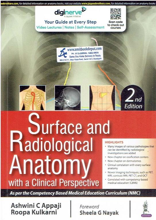
Surface and Radiological Anatomy with a Clinical Perspective, 2nd Edition, by Sheela G. Nayak, is the definitive guide for understanding human anatomy in a clinical context. Published by Jaypee Brothers, this richly illustrated book seamlessly integrates surface anatomy with modern imaging techniques. The book is divided into two sections, covering palpable bony landmarks and dermatomes, followed by an in-depth exploration of radiological anatomy, including CT, MRI, and contrast radiography procedures like barium studies and angiography. Designed for medical students and professionals, it bridges the gap between theoretical knowledge and practical application, making it an essential resource for exams and clinical practice.
Surface and Radiological Anatomy with a Clinical Perspective, 2nd Edition
Author: Sheela G. Nayak
Publisher: Jaypee Brothers Medical Publishers Pvt. Ltd.
A Foundational Text Bridging Anatomy and Clinical Practice
Mastering human anatomy is the cornerstone of medical education, but true clinical proficiency lies in the ability to translate two-dimensional diagrams into the three-dimensional living patient. Surface and Radiological Anatomy with a Clinical Perspective, 2nd Edition, by Sheela G. Nayak, serves as the essential guide for medical students and healthcare professionals seeking to bridge the gap between theoretical knowledge and practical application. This updated edition provides a comprehensive and visually rich exploration of anatomy as it is encountered in clinical examinations and modern diagnostic imaging.
The book is expertly structured into two complementary sections. The first section delves into surface anatomy, guiding the reader to identify and palpate critical bony landmarks, blood vessels, and nerves that lie beneath the skin. From the upper limb and lower limb to the complexities of the head and neck, this section ensures a solid grasp of the body’s visible and palpable structures. A dedicated chapter on dermatomes provides crucial insights into segmental nerve distribution, which is vital for neurological assessment.
The second, extensively revised section on radiological anatomy brings the subject into the modern era. It begins with the principles of radiology, offering clear explanations of core imaging modalities. Readers will gain a working knowledge of traditional radiography, contrast radiography, fluoroscopy, and mammography. The text then progresses to newer imaging techniques, demystifying complex technologies such as Computed Tomography (CT), Magnetic Resonance Imaging (MRI), Positron Emission Tomography (PET), and Doppler ultrasonography. By clearly outlining the differences between CT and MRI, the book equips students to understand the clinical rationale behind choosing specific imaging studies.
This foundational knowledge is then applied across all body regions. Chapters dedicated to the radiology of the upper limb, lower limb, thorax, abdomen and pelvis, and head and neck present images and line diagrams side-by-side for easy interpretation. Key clinical procedures are demystified through dedicated sections on contrast radiograms, including barium swallow, barium enema, cholecystography, endoscopic retrograde cholangiopancreatography (ERCP), pyelography, hysterosalpingography, carotid angiography, and coronary angiography. The book also features a comprehensive chapter on the ossification centers of the human skeletal system, a critical topic for understanding bone growth and development in pediatric radiology.
Key Features:
1. Integrated Approach: Seamlessly combines surface anatomy with its radiological correlation, providing a complete 3D understanding of the human body.
2. Clinical Perspective: Emphasizes the practical application of anatomical knowledge in patient examination and image interpretation, fostering clinical reasoning from the start.
3. Comprehensive Coverage: Includes detailed descriptions of both traditional radiographic views and modern cross-sectional imaging techniques like CT and MRI.
4. User-Friendly Format: Richly illustrated with high-quality diagrams and radiographs to facilitate visual learning and quick revision.
5. Exam-Oriented: An invaluable resource for preparing for practical examinations in anatomy and radiology.
Surface and Radiological Anatomy with a Clinical Perspective, 2nd Edition, is more than just a book; it is a clinical tool. Whether you are a first-year medical student learning to palpate bony landmarks or a resident refreshing your knowledge of magnetic resonance angiography, this book is an indispensable companion for your medical journey.
How does this book help a student prepare for spotting examinations in anatomy?
A1
The book is ideally structured for spotting exams. The first section provides clear descriptions and diagrams of surface bony landmarks and soft tissues. The second section offers high-quality radiographic images of all body regions, including the upper limb, thorax, and head and neck, with key features clearly labeled, making it perfect for identifying structures like the cardiac shadow or ossification centers.
Q2
Does the book cover advanced imaging techniques like MRI and CT scans?
A2
Yes, absolutely. Chapter 10, "Newer Imaging Techniques," is dedicated to explaining Computed Tomography (CT) , Magnetic Resonance Imaging (MRI) , and Positron Emission Tomography (PET) . It also provides a practical comparison of the differences between CT and MRI, helping students understand their respective clinical applications.
Q3
I struggle with identifying dermatomes. Is there a dedicated section for this?
A3
Yes, a full chapter (Chapter 7) is devoted to dermatomes. It provides a systematic overview of dermatomal distribution for the upper limb, lower limb, thorax, abdomen, and head and neck, which is fundamental knowledge for neurological examination.
Q4
Is there information on how to identify structures in an X-ray of the shoulder or knee?
A4
Certainly. Chapters 11 and 12 on the radiology of the upper limb and lower limb are specifically designed for this. They include detailed descriptions of normal radiographic anatomy for the shoulder region, elbow, hip region, knee joint, and ankle joint.
Q5
Does the book cover the anatomy of blood vessels as seen in angiograms?
A5
Yes, the book places a strong emphasis on vascular anatomy. It includes sections on coronary angiography, carotid angiography, vertebral artery angiography, and magnetic resonance angiography, correlating these images with the surface anatomy of the related blood vessels.
Q6
What is the "clinical perspective" mentioned in the title? Can you give an example?
A6
The clinical perspective is woven throughout the text. For example, when discussing the surface anatomy of the thyroid gland, it correlates its location with palpable landmarks in the neck. Similarly, in the radiology section, it explains the clinical significance of findings like an enlarged cardiac shadow or a narrowed vessel on an aortogram.
Q7
Does the book only cover bones, or are soft tissues and organs included?
A7
It provides comprehensive coverage of both. While bony landmarks are a key focus, the surface anatomy section also details the location of nerves. The radiology section extensively covers soft tissue shadows, such as the lung shadow, cardiac shadow, and mediastinal shadow in the thorax, and organs visualized through techniques like ultrasound and pyelography.
Q8
Does the book explain the basic physics behind how X-rays and MRIs work?
A8
Yes, the book starts with the principles of radiology in Chapter 9, defining key concepts like radiography, fluoroscopy, and tomography. It then explains the fundamental principles behind advanced techniques, including ultrasound, Doppler ultrasonography, and Magnetic Resonance Imaging.
Q9
Are there specific views for head and neck X-rays explained?
A9
Yes, Chapter 15 on the radiology of the head and neck is very thorough. It describes various standard views used in skull radiography, including the Posteroanterior (PA) view, Anteroposterior (AP) view, Lateral view, and the specialized Waters view for visualizing the facial bones and sinuses.
Q10
Besides medical students, who else would benefit from this book?
A10
This book is an excellent resource for a wide audience, including physiotherapy students learning to palpate bony landmarks; nursing and paramedical students; radiology technicians seeking a better understanding of the anatomy they image; and junior doctors as a quick refresher on clinically oriented anatomy.
0.00
0 Overall Rating
5
0
4
0
3
0
2
0
1
0
Try this product & share your review &
thoughts
SECTION 1: SURFACE ANATOMY
1. Introduction
- Anatomical Position of the Human Body
2. Surface Anatomy of Upper Limb
- Bony Landmarks
- Blood Vessels
- Nerves of the Upper Limb
3. Surface Anatomy of Lower Limb
- Bony Landmarks
4. Surface Anatomy of Thorax
- Bony and Soft Tissue Landmarks
5. Surface Anatomy of Head and Neck
- Regions of the Anterior Abdominal Wall
6. Surface Anatomy of Head and Neck
- Bony Landmarks
- Head and Neck
- Thyroid Gland
- Cranial Nerves
7. Dermatomes
- Dermatomes to the Upper Limb
- Dermatomes of the Lower Limb
- Dermatomes of the Thorax
- Dermatomes of the Abdomen
- Dermatomes of the Head and Neck
SECTION 2: RADIOLOGICAL ANATOMY
8. Introduction
9. Principles of Radiology
- Definition
- Radiography
- Radiographic Views
- Contrast Radiography
- Fluoroscopy
- Mammography
- Positron Emission Mammography
- Ultrasound
- 2D Ultrasound Images of Various Organs
- 3D and 4D Ultrasound
- Doppler Ultrasonography
10. Newer Imaging Techniques
- Tomography
- Computed Tomography/Computer Axial Tomography
- Positron Emission Tomography
- Magnetic Resonance Imaging or Nuclear Magnetic Resonance
- Differences Between CT and MRI
- Contrast MRI
- Magnetic Resonance Angiography
11. Radiology of Upper Limb
- Shoulder Region
- Elbow
- Wrist and Hand
12. Radiology of Lower Limb
- Hip Region
- Knee Joint
- Ankle Joint
- Foot
13. Radiology of Thorax
- Lung Shadow
- Cardiac Shadow
- Cardiac Size Estimation
- Mediastinal Shadow
- Skeletal Shadow
- Shadows of Diaphragm
- Contrast Radiograms Thoracic Region
- Bronchography
- Aortogram
- Barium Swallow
- Coronary Angiography
14. Radiology of Abdomen and Pelvis
- Contrast Radiogram of Abdomen and Pelvis
- Barium Enema
- Cholecystography
- Endoscopic Retrograde Cholangiopancreatography
- Pyelography
- Hysterosalpingography
15. Radiology of Head and Neck
- Views
- Posteroanterior View
- Anteroposterior View
- Lateral View
- Superior View
- Waters View
- Radiology of Neck
- Carotid Angiography
- Vertebral Artery Angiography
- Sialography
16. Ossification Centers of the Human Skeletal System
Surface and Radiological Anatomy with a Clinical Perspective, 2nd Edition
Author: Sheela G. Nayak
Publisher: Jaypee Brothers Medical Publishers Pvt. Ltd.
A Foundational Text Bridging Anatomy and Clinical Practice
Mastering human anatomy is the cornerstone of medical education, but true clinical proficiency lies in the ability to translate two-dimensional diagrams into the three-dimensional living patient. Surface and Radiological Anatomy with a Clinical Perspective, 2nd Edition, by Sheela G. Nayak, serves as the essential guide for medical students and healthcare professionals seeking to bridge the gap between theoretical knowledge and practical application. This updated edition provides a comprehensive and visually rich exploration of anatomy as it is encountered in clinical examinations and modern diagnostic imaging.
The book is expertly structured into two complementary sections. The first section delves into surface anatomy, guiding the reader to identify and palpate critical bony landmarks, blood vessels, and nerves that lie beneath the skin. From the upper limb and lower limb to the complexities of the head and neck, this section ensures a solid grasp of the body’s visible and palpable structures. A dedicated chapter on dermatomes provides crucial insights into segmental nerve distribution, which is vital for neurological assessment.
The second, extensively revised section on radiological anatomy brings the subject into the modern era. It begins with the principles of radiology, offering clear explanations of core imaging modalities. Readers will gain a working knowledge of traditional radiography, contrast radiography, fluoroscopy, and mammography. The text then progresses to newer imaging techniques, demystifying complex technologies such as Computed Tomography (CT), Magnetic Resonance Imaging (MRI), Positron Emission Tomography (PET), and Doppler ultrasonography. By clearly outlining the differences between CT and MRI, the book equips students to understand the clinical rationale behind choosing specific imaging studies.
This foundational knowledge is then applied across all body regions. Chapters dedicated to the radiology of the upper limb, lower limb, thorax, abdomen and pelvis, and head and neck present images and line diagrams side-by-side for easy interpretation. Key clinical procedures are demystified through dedicated sections on contrast radiograms, including barium swallow, barium enema, cholecystography, endoscopic retrograde cholangiopancreatography (ERCP), pyelography, hysterosalpingography, carotid angiography, and coronary angiography. The book also features a comprehensive chapter on the ossification centers of the human skeletal system, a critical topic for understanding bone growth and development in pediatric radiology.
Key Features:
1. Integrated Approach: Seamlessly combines surface anatomy with its radiological correlation, providing a complete 3D understanding of the human body.
2. Clinical Perspective: Emphasizes the practical application of anatomical knowledge in patient examination and image interpretation, fostering clinical reasoning from the start.
3. Comprehensive Coverage: Includes detailed descriptions of both traditional radiographic views and modern cross-sectional imaging techniques like CT and MRI.
4. User-Friendly Format: Richly illustrated with high-quality diagrams and radiographs to facilitate visual learning and quick revision.
5. Exam-Oriented: An invaluable resource for preparing for practical examinations in anatomy and radiology.
Surface and Radiological Anatomy with a Clinical Perspective, 2nd Edition, is more than just a book; it is a clinical tool. Whether you are a first-year medical student learning to palpate bony landmarks or a resident refreshing your knowledge of magnetic resonance angiography, this book is an indispensable companion for your medical journey.
SECTION 1: SURFACE ANATOMY
1. Introduction
- Anatomical Position of the Human Body
2. Surface Anatomy of Upper Limb
- Bony Landmarks
- Blood Vessels
- Nerves of the Upper Limb
3. Surface Anatomy of Lower Limb
- Bony Landmarks
4. Surface Anatomy of Thorax
- Bony and Soft Tissue Landmarks
5. Surface Anatomy of Head and Neck
- Regions of the Anterior Abdominal Wall
6. Surface Anatomy of Head and Neck
- Bony Landmarks
- Head and Neck
- Thyroid Gland
- Cranial Nerves
7. Dermatomes
- Dermatomes to the Upper Limb
- Dermatomes of the Lower Limb
- Dermatomes of the Thorax
- Dermatomes of the Abdomen
- Dermatomes of the Head and Neck
SECTION 2: RADIOLOGICAL ANATOMY
8. Introduction
9. Principles of Radiology
- Definition
- Radiography
- Radiographic Views
- Contrast Radiography
- Fluoroscopy
- Mammography
- Positron Emission Mammography
- Ultrasound
- 2D Ultrasound Images of Various Organs
- 3D and 4D Ultrasound
- Doppler Ultrasonography
10. Newer Imaging Techniques
- Tomography
- Computed Tomography/Computer Axial Tomography
- Positron Emission Tomography
- Magnetic Resonance Imaging or Nuclear Magnetic Resonance
- Differences Between CT and MRI
- Contrast MRI
- Magnetic Resonance Angiography
11. Radiology of Upper Limb
- Shoulder Region
- Elbow
- Wrist and Hand
12. Radiology of Lower Limb
- Hip Region
- Knee Joint
- Ankle Joint
- Foot
13. Radiology of Thorax
- Lung Shadow
- Cardiac Shadow
- Cardiac Size Estimation
- Mediastinal Shadow
- Skeletal Shadow
- Shadows of Diaphragm
- Contrast Radiograms Thoracic Region
- Bronchography
- Aortogram
- Barium Swallow
- Coronary Angiography
14. Radiology of Abdomen and Pelvis
- Contrast Radiogram of Abdomen and Pelvis
- Barium Enema
- Cholecystography
- Endoscopic Retrograde Cholangiopancreatography
- Pyelography
- Hysterosalpingography
15. Radiology of Head and Neck
- Views
- Posteroanterior View
- Anteroposterior View
- Lateral View
- Superior View
- Waters View
- Radiology of Neck
- Carotid Angiography
- Vertebral Artery Angiography
- Sialography
16. Ossification Centers of the Human Skeletal System
How does this book help a student prepare for spotting examinations in anatomy?
A1
The book is ideally structured for spotting exams. The first section provides clear descriptions and diagrams of surface bony landmarks and soft tissues. The second section offers high-quality radiographic images of all body regions, including the upper limb, thorax, and head and neck, with key features clearly labeled, making it perfect for identifying structures like the cardiac shadow or ossification centers.
Q2
Does the book cover advanced imaging techniques like MRI and CT scans?
A2
Yes, absolutely. Chapter 10, "Newer Imaging Techniques," is dedicated to explaining Computed Tomography (CT) , Magnetic Resonance Imaging (MRI) , and Positron Emission Tomography (PET) . It also provides a practical comparison of the differences between CT and MRI, helping students understand their respective clinical applications.
Q3
I struggle with identifying dermatomes. Is there a dedicated section for this?
A3
Yes, a full chapter (Chapter 7) is devoted to dermatomes. It provides a systematic overview of dermatomal distribution for the upper limb, lower limb, thorax, abdomen, and head and neck, which is fundamental knowledge for neurological examination.
Q4
Is there information on how to identify structures in an X-ray of the shoulder or knee?
A4
Certainly. Chapters 11 and 12 on the radiology of the upper limb and lower limb are specifically designed for this. They include detailed descriptions of normal radiographic anatomy for the shoulder region, elbow, hip region, knee joint, and ankle joint.
Q5
Does the book cover the anatomy of blood vessels as seen in angiograms?
A5
Yes, the book places a strong emphasis on vascular anatomy. It includes sections on coronary angiography, carotid angiography, vertebral artery angiography, and magnetic resonance angiography, correlating these images with the surface anatomy of the related blood vessels.
Q6
What is the "clinical perspective" mentioned in the title? Can you give an example?
A6
The clinical perspective is woven throughout the text. For example, when discussing the surface anatomy of the thyroid gland, it correlates its location with palpable landmarks in the neck. Similarly, in the radiology section, it explains the clinical significance of findings like an enlarged cardiac shadow or a narrowed vessel on an aortogram.
Q7
Does the book only cover bones, or are soft tissues and organs included?
A7
It provides comprehensive coverage of both. While bony landmarks are a key focus, the surface anatomy section also details the location of nerves. The radiology section extensively covers soft tissue shadows, such as the lung shadow, cardiac shadow, and mediastinal shadow in the thorax, and organs visualized through techniques like ultrasound and pyelography.
Q8
Does the book explain the basic physics behind how X-rays and MRIs work?
A8
Yes, the book starts with the principles of radiology in Chapter 9, defining key concepts like radiography, fluoroscopy, and tomography. It then explains the fundamental principles behind advanced techniques, including ultrasound, Doppler ultrasonography, and Magnetic Resonance Imaging.
Q9
Are there specific views for head and neck X-rays explained?
A9
Yes, Chapter 15 on the radiology of the head and neck is very thorough. It describes various standard views used in skull radiography, including the Posteroanterior (PA) view, Anteroposterior (AP) view, Lateral view, and the specialized Waters view for visualizing the facial bones and sinuses.
Q10
Besides medical students, who else would benefit from this book?
A10
This book is an excellent resource for a wide audience, including physiotherapy students learning to palpate bony landmarks; nursing and paramedical students; radiology technicians seeking a better understanding of the anatomy they image; and junior doctors as a quick refresher on clinically oriented anatomy.
No Syllabus Added
0.00
0 Overall Rating
5
0
4
0
3
0
2
0
1
0
Try this product & share your review &
thoughts
Top Trending Product
Related
Product
Related Product
Related Blog Posts
Latest Blogs
Latest Blogs
Classic Literature Reimagined: Discuss modern twists on classic novels.
Lorem ipsum dolor sit amet, consectetur adipiscing elit, sed
do
eiusmod tempor incididunt ut labore et dolore magna aliqua. Ut enim ad minim
veniam, quis nostrud exercitation ullamco Lorem ipsum dolor sit amet,
consectetur adipiscing elit, sed do eiusmod tempor incididunt ut labore et
dolore magna aliqua. Utenim ad minim veniam, quis nostrud exercitation
ullamco
Lorem ipsum dolor sit amet, consecte...